Peritoneal Mesothelioma Ct Scan / A Novel Prognostic Model For Malignant Mesothelioma Incorporating Quantitative Fdg Pet Imaging With Clinical Parameters Clinical Cancer Research / The journal of thoracic disease indicates that not only does mesothelioma show up on a ct scan, but it is the preferred diagnostic tool of choice for advanced stage mesothelioma cases.
Peritoneal Mesothelioma Ct Scan / A Novel Prognostic Model For Malignant Mesothelioma Incorporating Quantitative Fdg Pet Imaging With Clinical Parameters Clinical Cancer Research / The journal of thoracic disease indicates that not only does mesothelioma show up on a ct scan, but it is the preferred diagnostic tool of choice for advanced stage mesothelioma cases.. Because adequate cytoreduction is necessary to achieve prolonged survival, ct scans became an accurate prognostic radiologic test for patient selection for comprehensive treatment. Sometimes, a repeat biopsy is required and may need to be reviewed by a pathologist with expertise in mesothelioma. peritoneal mesothelioma 131 the case reported by dach et al. It is seen in women with prior gynaecological surgery or infection. Diaphragmatic invasion, ascites, and omental caking are common ct scan findings of peritoneal mesothelioma.
ct scan can also play an important role in guiding biopsy for tissue diagnosis and can provide the surgeon with a A biopsy is always required for confirmation. Kebapci m, vardareli e, adapinar b, et al. Malignant peritoneal mesothelioma (mpm) is a rare but fatal tumor. Many factors are suspected to contribute to its development, such as previous surgery, endometriosis, and familial mediterranean fever.
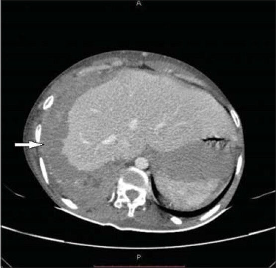
Extraperitoneal Parastomal Metastasis Of Malignant Peritoneal Mesothelioma Bmj Case Reports from casereports.bmj.com These imaging studies are done to assess the extent of disease in the abdomen. The clinical presentations and imaging findings are nonspecific and resemble various diseases, including peritoneal metastasis. Worrisome lesions seen on ct imaging would prompt a biopsy attempt to confirm the diagnosis. Doctors use ct scans to determine if a tumor has spread to the chest wall or diaphragm. It can help identify the best therapies to control the cancer while maximizing quality. Benign multicystic peritoneal mesothelioma (bmpm) is a rare condition that arises from the abdominal peritoneum. ct scans and mris aren't used as often. The peritoneum is a preferred site of metastasis of several primary
A ct scan can show abnormal swellings in the lining of your lungs (pleura) or abdomen (peritoneum).
This name is very confusing since there is no relation with the malignant mesothelioma. Both patients had ct scans of the abdomen. Bmpm usually affects premenopausal women and is extremely rare in men. Pet/ct scan challenge of pleural effusion treatment for mesothelioma patients. The peritoneum is a preferred site of metastasis of several primary Worrisome lesions seen on ct imaging would prompt a biopsy attempt to confirm the diagnosis. mesothelioma tests commonly include imaging scans, such as mri and ct scans, and biopsies such as pleural aspiration or thoracoscopy. Suggest mesothelioma include an abnormal thickening of the pleura, calcium deposits on the pleura, fluid in the space between the lungs and the chest wall, or changes in the lungs themselves as a result of asbestos exposure. However, the exact diagnosis and characterization of lesions may be diffi cult due to the overlap of imaging fi ndings. Your gp will conduct a physical examination and order tests. Blood and urine analysis may be ordered to check for signs indicative of cancer, such as elevated white blood cells or proteins. Report of 11 new cases and review of the literature. peritoneal mesothelioma is a rare cancer of the abdominal lining with about 600 cases per year in the united states.
A ct scan is approximately 90% sensitive for detecting malignant pleural mesothelioma. The images are taken from numerous angles so that doctors have a full view. For example, an appendix cancer could be diagnosed during what would have been a routine appendectomy. Exposure to asbestos fibers can cause mesothelioma even years later. It is usually associated with asbestos exposure and regarded as universally fatal.
Malignant Pleural And Peritoneal Mesothelioma Consequential To Brief Indirect Asbestos Exposure Journal Of Clinical Imaging Science from clinicalimagingscience.org Suggest mesothelioma include an abnormal thickening of the pleura, calcium deposits on the pleura, fluid in the space between the lungs and the chest wall, or changes in the lungs themselves as a result of asbestos exposure. A biopsy is the only definitive way to confirm a mesothelioma diagnosis. ct scans performed in 53 patients with mpm and 27 patients with tbp confirmed by pathology were retrospectively reviewed. The results were correlated with either contemporaneous peritoneal biopsy or ascitic aspirate or with radiographic or. mesothelioma refers to a tumor in the lining of the lung, stomach, heart, or other organs that. Computerized tomography scan (ct scan) helps determine the location, size and extent of mesothelioma tumors and can help determine whether the tumor has invaded any of the adjacent structures. Benign multicystic peritoneal mesothelioma (bmpm) is a rare condition that arises from the abdominal peritoneum. Because adequate cytoreduction is necessary to achieve prolonged survival, ct scans became an accurate prognostic radiologic test for patient selection for comprehensive treatment.
ct scan can also play an important role in guiding biopsy for tissue diagnosis and can provide the surgeon with a
However, the exact diagnosis and characterization of lesions may be diffi cult due to the overlap of imaging fi ndings. The second patient was also examined with mri. ct scans performed in 53 patients with mpm and 27 patients with tbp confirmed by pathology were retrospectively reviewed. Secondary peritoneal malignances are by far much more common than primary peritoneal tumors. The peritoneum is a preferred site of metastasis of several primary According to the national cancer institute, the peritoneum is the layer of tissue that lines the entire abdominal wall and essentially covers all of the. However, they can help the doctor understand the extent and stage of disease. A biopsy is always required for confirmation. Diagnosis of peritoneal mesothelioma is usually suspected when someone seeks medical attention and a diagnostic exam, such as a ct scan or an ultrasound of the abdomen, shows masses or fluid in. Worrisome lesions seen on ct imaging would prompt a biopsy attempt to confirm the diagnosis. Doctors also use mesothelioma blood tests to measure treatment response. ct scans and mris for mesothelioma diagnosis. Common primary peritoneal tumor is mesothelioma.
The results were correlated with either contemporaneous peritoneal biopsy or ascitic aspirate or with radiographic or. ct scans performed in 53 patients with mpm and 27 patients with tbp confirmed by pathology were retrospectively reviewed. A recent report of anteriography in peritoneal mesothelioma described three cases of mildly to moderately hypervascular omental lesions without arteriovenous shunting; The aim of this study was to determine which computed tomography (ct) findings were useful in differentiating malignant peritoneal mesothelioma (mpm) and tuberculous peritonitis (tbp). peritoneal mesothelioma your guide to best cancer care.
Pseudomyxoma Peritonei from ct scans and mris for mesothelioma diagnosis. The ct scan supplies correct details about the situation and thickness of the tumour(s) within the chest or stomach. Patients have either a ct scan or mri of the abdomen and pelvis to confirm the presence of new or recurrent disease in the peritoneum. A biopsy is the only definitive way to confirm a mesothelioma diagnosis. ct shows an increased soft report of us and ct demonstration of scalloping of tissue density affecting the omentum, a diffuse or a liver edge in a patient of peritoneal mesothelioma. A ct scan may be performed in order to diagnose peritoneal mesothelioma. peritoneal mesothelioma is a rare cancer of the abdominal lining with about 600 cases per year in the united states. How is peritoneal disease diagnosed?
Blood and urine analysis may be ordered to check for signs indicative of cancer, such as elevated white blood cells or proteins.
Radiology imaging sometimes will not pick up evidence of diffuse cancerous growths on the mesothelium, but will readily produce a shadow created by a single tumor that may occur more readily with peritoneal mesothelioma. ct scan plays an important role in the detection of peritoneal carcinomatosis and its mimics. Diagnosis how is peritoneal mesothelioma diagnosed? ct scan can also play an important role in guiding biopsy for tissue diagnosis and can provide the surgeon with a Malignant peritoneal mesothelioma is a rare tumor with a variable appearance at ct 1. The aim of the current study was to identify computed tomography (ct) scan images that are useful in patient selection for this comprehensive approach. The clinical presentations and imaging findings are nonspecific and resemble various diseases, including peritoneal metastasis. A ct scan may suggest the presence of cancer in the abdominal cavity, but cannot confirm the diagnosis of mpm. The data gathered by the ct scan is used to work out one of the simplest ways of acquiring tissue for testing (see biopsy beneath). It might probably additionally present if the mesothelioma has unfolded to different organs. Diagnosis of peritoneal mesothelioma is usually suspected when someone seeks medical attention and a diagnostic exam, such as a ct scan or an ultrasound of the abdomen, shows masses or fluid in. peritoneal mesothelioma your guide to best cancer care. Research from ut southwestern medical center indicates that peritoneal mesothelioma can spread to other organs, such as the lungs, in the last and most serious stage of this cancer, stage 4a or stage 4b.


0 Comments